
Cognizin® delivers a patented form of citicoline. Citicoline is a brain health ingredient that provides support for attention, focus and recall. Clinical studies have shown Cognizin to help improve attention, focus, motor speed and increase brain activity. †
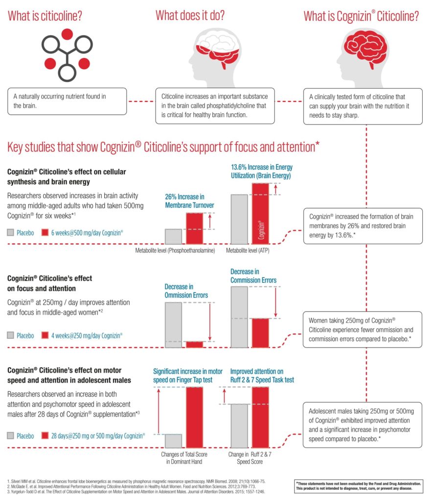
Results: These findings indicate that long-term dietary CDP-choline supplementation can ameliorate the hippocampal-dependent memory impairment caused by impoverished environmental conditions in rats, and suggest that its actions result, in part, from a long-term effect such as enhanced membrane phosphatide synthesis, an effect shown to require long-term dietary supplementation with CDP-choline.
Learn Mem. 2005 Jan-Feb;12(1):39-43. Epub 2005 Jan 12.
Teather LA1, Wurtman RJ.
http://www.learnmem.org/cgi/doi/10.1101/lm.83905
Results: Overall, these findings suggest that the beverage significantly improved sustained attention, cognitive effort and reaction times in healthy adults. Evidence of improved P450 amplitude indicates a general improvement in the ability to accommodate new and relevant information within working memory and overall enhanced brain activation.
Int J Food Sci Nutr. 2014 Dec;65(8):1003-7. doi: 10.3109/09637486.2014.940286. Epub 2014 Jul 21.
Bruce SE1, Werner KB, Preston BF, Baker LM.
https://doi.org/10.3109/09637486.2014.940286
Results: This is the first study to report a direct effect of uridine on membrane phospholipid precursors in healthy adults using (31) P-MRS. Sustained administration of uridine appears to increase PME in healthy subjects. Further investigation is required to clarify the effects of uridine in disorders with altered phospholipid metabolism such as bipolar disorder.
Bipolar Disord. 2010 Dec;12(8):825-33. doi: 10.1111/j.1399-5618.2010.00884.x.
Agarwal N1, Sung YH, Jensen JE, daCunha G, Harper D, Olson D, Renshaw PF.
https://doi.org/10.1111/j.1399-5618.2010.00884.x
An amino acid that is naturally produced in the body which helps produce energy and can help increase energy production in the mitochondria. This acetylated version of carnitine is able to cross the blood-brain barrier. Its primary effect is on the production of acetylcholine which is one of the main neurotransmitters involved in memory, learning, computation, analysis and many other cognitive processes.†
Results: Overall, our findings indicate a systemic positive effect of ALCAR dietary treatment and a tissue specific regulation of mitochondrial homeostasis in brain of old rats. Moreover, it appears that ALCAR acts as a nutrient since in most cases its effects were almost completely abolished one month after treatment suspension. Dietary supplementation of old rats with this compound seems a valuable approach to prevent age-related mitochondrial dysfunction and might ultimately represent a strategy to delay age-associated negative consequences in mitochondrial homeostasis.
Exp Gerontol. 2017 Nov;98:99-109. doi: 10.1016/j.exger.2017.08.017. Epub 2017 Aug 12.
Nicassio L1, Fracasso F1, Sirago G1, Musicco C2, Picca A3, Marzetti E3, Calvani R3, Cantatore P1, Gadaleta MN2, Pesce V4.
https://doi.org/10.1016/j.exger.2017.08.017
Results: There is compelling evidence from preclinical studies that L-carnitine and ALCAR can improve energy status, decrease oxidative stress and prevent subsequent cell death in models of adult, neonatal and pediatric brain injury. ALCAR can provide an acetyl moiety that can be oxidized for energy, used as a precursor for acetylcholine, or incorporated into glutamate, glutamine and GABA, or into lipids for myelination and cell growth. Administration of ALCAR after brain injury in rat pups improved long-term functional outcomes, including memory. Additional studies are needed to better explore the potential of L-carnitine and ALCAR for protection of developing brain as there is an urgent need for therapies that can improve outcome after neonatal and pediatric brain injury.
Neurochem Res. 2017 Jun;42(6):1661-1675. doi: 10.1007/s11064-017-2288-7. Epub 2017 May 16.
Ferreira GC1,2, McKenna MC3,4.
https://link.springer.com/article/10.1007%2Fs11064-017-2288-7
Results: The results of this study clearly suggest the neuroprotective effect of ALC in 6-OHDA-induced model of PD through abrogation of neuroinflammation, apoptosis, astrogliosis, and oxidative stress and it may be put forward as an ancillary therapeutic candidate for controlling PD.
Biomed Pharmacother. 2017 May;89:1-9. doi: 10.1016/j.biopha.2017.02.007. Epub 2017 Feb 12.
Afshin-Majd S1, Bashiri K2, Kiasalari Z3, Baluchnejadmojarad T4, Sedaghat R5, Roghani M6.
https://doi.org/10.1016/j.biopha.2017.02.007
Results: These results have been discussed considering the pathophysiology and treatment of Dementias and Depression because, although referred to normal healthy animals, they support the notion that l-acetylcarnitine may have positive effects in these pathologies.
Eur J Pharmacol. 2015 Jun 5;756:67-74. doi: 10.1016/j.ejphar.2015.03.011. Epub 2015 Mar 19.
Ferrari F1, Gorini A1, Villa RF2
https://doi.org/10.1016/j.ejphar.2015.03.011
Results: These findings are consistent with decreased metabolism of glucose to lactate but not via the TCA cycle. Higher amounts of the sum of adenosine nucleotides, phosphocreatine and the phosphocreatine/creatine ratio found in the cortex of ALCAR-treated mice are indicative of increased energy levels. Furthermore, ALCAR supplementation increased the levels of the neurotransmitters noradrenaline in the HF and serotonin in cortex, consistent with ALCAR’s potential efficacy for depressive symptoms. Other ALCAR-induced changes observed included reduced amounts of GABA in the HF and increased myo-inositol. In conclusion, chronic ALCAR supplementation decreased glucose metabolism to lactate, resulted in increased energy metabolite and altered monoamine neurotransmitter levels in the mouse brain.
Neurochem Int. 2012 Jul;61(1):100-7. doi: 10.1016/j.neuint.2012.04.008. Epub 2012 Apr 23.
Smeland OB1, Meisingset TW, Borges K, Sonnewald U.
https://doi.org/10.1016/j.neuint.2012.04.008

Hericium Erinaceus (also called Lion’s Mane) is an edible and medicinal mushroom. Lion’s Mane can induce Nerve Growth Factor synthesis in nerve cells which is essential for the maintenance of the basal forebrain cholinergic system. A double-blind placebo-controlled clinical trial study showed increased cognitive function scores for people who took Lion’s Mane vs a placebo group. †
Results: The extract contained neuroactive compounds that induced the secretion of extracellular NGF in NG108-15 cells, thereby promoting neurite outgrowth activity. However, the H. erinaceus extract failed to protect NG108-15 cells subjected to oxidative stress when applied in pre-treatment and co-treatment modes. In conclusion, the aqueous extract of H. erinaceus contained neuroactive compounds which induced NGF-synthesis and promoted neurite outgrowth in NG108-15 cells. The extract also enhanced the neurite outgrowth stimulation activity of NGF when applied in combination. The aqueous preparation of H. erinaceus had neurotrophic but not neuroprotective activities.
Int J Med Mushrooms. 2013;15(6):539-54.
Lai PL1, Naidu M, Sabaratnam V, Wong KH, David RP, Kuppusamy UR, Abdullah N, Malek SN.
Evid Based Complement Alternat Med. 2017;2017:3864340. doi: 10.1155/2017/3864340. Epub 2017 Jan 1.
Brandalise F1, Cesaroni V2, Gregori A3, Repetti M2, Romano C2, Orrù G4, Botta L2, Girometta C5, Guglielminetti ML6, Savino E6, Rossi P7.
Results: Intriguingly other neurobiological effects have recently been reported like the effect on neurite outgrowth and differentiation in PC12 cells. Until now no investigations have been conducted to assess the impact of this dietary supplementation on brain function in healthy subjects. Therefore, we have faced the problem by considering the effect on cognitive skills and on hippocampal neurotransmission in wild-type mice. In wild-type mice the oral supplementation with H. erinaceus induces, in behaviour test, a significant improvement in the recognition memory and, in hippocampal slices, an increase in spontaneous and evoked excitatory synaptic current in mossy fiber-CA3 synapse. In conclusion, we have produced a series of findings in support of the concept that H. erinaceus induces a boost effect onto neuronal functions also in non-pathological conditions.
https://doi.org/10.1155/2017/3864340
Results: The results obtained in this study suggest that Yamabushitake is effective in improving mild cognitive impairment.
Phytother Res. 2009 Mar;23(3):367-72. doi: 10.1002/ptr.2634.
Mori K1, Inatomi S, Ouchi K, Azumi Y, Tuchida T.
https://doi.org/10.1002/ptr.2634
Results: Our results show that HE intake has the possibility to reduce depression and anxiety and these results suggest a different mechanism from NGF-enhancing action of H. erinaceus.
Biomed Res. 2010 Aug;31(4):231-7.
Nagano M1, Shimizu K, Kondo R, Hayashi C, Sato D, Kitagawa K, Ohnuki K.

Rhodiola rosea is a perennial flowering plant in the family Crassulaceae. In some clinical trials Rhodiola Rosea has been found to show anti fatiguing benefits for brain function.†
Results: Our results showed that a single i.p. administration of salidroside was able to enhance fear memory and exerted an anxiolytic and antidepressant effect. These data confirmed the adaptogenic effect of R. Rosea bioactive compounds in animal models and suggest that salidroside might represent an interesting pharmacological tool to ameliorate cognition and counteract mood disorders.
J Alzheimers Dis.2016 Feb 26;52(1):65-75. doi: 10.3233/JAD-151159.
Palmeri A, Mammana L, Tropea MR, Gulisano W, Puzzo D.
https://doi.org/10.3233/JAD-151159
Results: The chemical fingerprinting of the standardized commercial Rhodiola extract was performed by means of nuclear magnetic resonance (NMR). Naive rats treated with standardized Rhodiola extract increased the number of avoidances during the learning session and memory retention test compared to the controls. Rats with scopolamine-induced memory impairment treated with Rhodiola extract showed an increase in the number of avoidances during the learning session and on the memory tests compared to the scopolamine group. The other two parameters were not changed in rats treated with the extract of Rhodiola in the two series.
J Ethnopharmacol. 2016 Dec 4;193:586-591. doi: 10.1016/j.jep.2016.10.011. Epub 2016 Oct 5.
Vasileva LV1, Getova DP2, Doncheva ND3, Marchev AS4, Georgiev MI4.
https://doi.org/10.1016/j.jep.2016.10.011
Results: Our results show that HE intake has the possibility to reduce depression and anxiety and these results suggest a different mechanism from NGF-enhancing action of H. erinaceus.
Biomed Res. 2010 Aug;31(4):231-7.
Nagano M1, Shimizu K, Kondo R, Hayashi C, Sato D, Kitagawa K, Ohnuki K.
https://doi.org/10.1016/j.jep.2016.10.011
Results: The efficacy of SHR-5 extract with respect to depressive complaints was assessed on days 0 and 42 of the study period from total and specific subgroup HAMD scores. For individuals in groups A and B, overall depression, together with insomnia, emotional instability and somatization, but not self-esteem, improved significantly following medication, whilst the placebo group did not show such improvements. No serious side-effects were reported in any of the groups A–C.
Nord J Psychiatry. 2007;61(5):343-8.
Darbinyan V1, Aslanyan G, Amroyan E, Gabrielyan E, Malmström C, Panossian A.
https://doi.org/10.1080/08039480701643290
Results: The MDA level was significantly increased while the GR and GSH levels were significantly decreased with striking impairments in spatial learning and memory and severe damage to hippocampal neurons in the model rat induced by intracerebroventricular injection of streptozotocin. These abnormalities were significantly improved by pretreatment with R. rosea extract (3.0 g/kg).
Biomed Environ Sci. 2009 Aug;22(4):318-26. doi: 10.1016/S0895-3988(09)60062-3.
Qu ZQ1, Zhou Y, Zeng YS, Li Y, Chung P.
https://doi.org/10.1016/S0895-3988(09)60062-3
Curcumin is a natural phenolic component of yellow curry spice, which is used in some cultures for the treatment of diseases associated with oxidative stress and inflammation. Recent studies show that curcumin also possesses neuroprotective and cognitive-enhancing properties.†
Results: Suggesting an overall improvement in spatial attention. Measures of visual discrimination learning, executive function and spatial memory, and levels of brain and cerebrospinal fluid Aβ were unaffected by the cocktail. Our results indicate that this medical food cocktail may be beneficial for improving spatial attention and motivation deficits associated with impaired cognition in aging and AD.
J Alzheimers Dis. 2012;32(4):1029-42. doi: 10.3233/JAD-2012-120937.
Head E1, Murphey HL, Dowling AL, McCarty KL, Bethel SR, Nitz JA, Pleiss M, Vanrooyen J, Grossheim M, Smiley JR, Murphy MP, Beckett TL, Pagani D, Bresch F, Hendrix C.
https://doi.org/10.3233/JAD-2012-120937
Results: These compounds salvaged PSD-95, synaptophysin and camkIV expression levels in the hippocampus in the rat AD model, which suggests multiple target sites with the potential of curcuminoids in spatial memory enhancing and disease modifying in AD.
Neuroscience. 2010 Sep 1;169(3):1296-306. doi: 10.1016/j.neuroscience.2010.05.078. Epub 2010 Jun 9
Ahmed T1, Enam SA, Gilani AH.
https://doi.org/10.1016/j.neuroscience.2010.05.078
Results: Six clinical trials with a total of 377 patients were reviewed, comparing the use of curcumin to placebo. In patients with depression, the pooled standardized mean difference from baseline Hamilton Rating Scale for Depression scores (pooled standardized mean difference −0.344, 95% confidence interval −0.558 to −0.129; P = .002) support the significant clinical efficacy of curcumin in ameliorating depressive symptoms. Significant anti-anxiety effects were also reported in 3 of the trials. Notably, no adverse events were reported in any of the trials. Most trials had a generally low risk of bias, except for an open trial of curcumin and a single-blinded study.
J Am Med Dir Assoc. 2017 Jun 1;18(6):503-508. doi: 10.1016/j.jamda.2016.12.071. Epub 2017 Feb 22.
Ng QX1, Koh SSH2, Chan HW3, Ho CYX3.
https://doi.org/10.1016/j.neuroscience.2010.05.078
Results: Ar-turmerone, curcuminoids and α-linolenic acid were the lead compounds present in turmeric extract. Increased serum corticosterone levels were observed in stressed mice when compared to the control group, while turmeric treatment significantly reduced serum corticosterone level. Turmeric treatment caused an improved learning and memory in Morris water maze test in stressed animals. Social novelty preference was also restored in turmeric treated animals. Following turmeric treatment, M5 expression was improved in the cortex and M3 expression was improved in the hippocampus of stress + scopolamine + turmeric treated group.
Curr Drug Targets. 2017;18(13):1545-1557. doi: 10.2174/1389450118666170315120627.
Khalid A1, Shakeel R1, Justin S1, Iqbal G1, Shah SAA2, Zahid S1, Ahmed T1.
https://doi.org/10.2174/1389450118666170315120627
Results: The findings therefore suggest that supplementation of curcumin may be beneficial in the treatment of acute stress induced anxiety and enhancement of memory function.
Brain Res Bull. 2015 Jun;115:1-8. doi: 10.1016/j.brainresbull.2015.04.001. Epub 2015 Apr 11.
Haider S1, Naqvi F2, Batool Z3, Tabassum S4, Sadir S2, Liaquat L2, Naqvi F2, Zuberi NA5, Shakeel H2, Perveen T2.
https://doi.org/10.1016/j.brainresbull.2015.04.001

Phosphatidylserine is a phospholipid and is a component of the cell membrane. It plays a key role in cell cycle signaling, specifically in relationship to apoptosis. In a recent Japenese study, with oral administration of Soy-PS for 6 months improved memory function, especially delayed recall, in the elderly with memory complaints. †
Results: PS is efficiently absorbed after oral consumption. A positive influence of PS+PA on memory, mood, and cognition was demonstrated among elderly test subjects. Short-term supplementation with PS+PA in patients with AD showed a stabilizing effect on daily functioning, emotional state and self-reported general condition. The data encourage long-term studies with PS+PA in AD patients and other elderly with memory or cognition problems.
Adv Ther.2014 Dec;31(12):1247-62. doi: 10.1007/s12325-014-0165-1. Epub 2014 Nov 21.
Moré MI1, Freitas U, Rutenberg D.
https://doi.org/10.1007/s12325-014-0165-1
Results: As a result, there was no difference in blood markers and vital signs during Soy-PS treatment and any side effect caused by Soy-PS treatment was not observed. Neuropsychological test scores were similarly increased in all groups including placebo group. However, in the subjects with relatively low score at baseline, the memory scores in PS treated groups were significantly increased against the baseline, while those of placebo group remained unchanged. And the memory improvements in Soy-PS-treated groups were mostly attributed to the increase in delayed verbal recall, a memory ability attenuated in the earliest stage of dementia. In conclusion, Soy-PS used in this study is considered as safety food ingredient and 6 months of Soy-PS supplementation could improve the memory functions of the elderly with memory complaints.
J Clin Biochem Nutr. 2010 Nov;47(3):246-55. doi: 10.3164/jcbn.10-62. Epub 2010 Sep 29.
Kato-Kataoka A1, Sakai M, Ebina R, Nonaka C, Asano T, Miyamori T.
https://doi.org/10.3164/jcbn.10-62
Results: Chronic stress levels and other baseline measures did not differ between treatment groups (all p > 0.05). Acute stress was successfully induced by the TSST and resulted in a hyper-responsivity of the HPAA in chronically stressed subjects. Compared to placebo, a supplementation with a daily dose of PAS 400 was effective in normalizing the ACTH (p = 0.010), salivary (p = 0.043) and serum cortisol responses (p = 0.035) to the TSST in chronically high but not in low stressed subjects (all p > 0.05). Compared to placebo, supplementation with PAS 200 did not result in any significant differences in these variables (all p > 0.05). There were no significant effects of supplementation with PAS on heart rate, pulse transit time, or psychological stress response (all p > 0.05).
Lipids Health Dis. 2014 Jul 31;13:121. doi: 10.1186/1476-511X-13-121.
Hellhammer J1, Vogt D, Franz N, Freitas U, Rutenberg D.
https://doi.org/10.1186/1476-511X-13-121
Results: Treatment with 400 mg PAS resulted in a pronounced blunting of both serum ACTH and cortisol, and salivary cortisol responses to the TSST, but did not affect heart rate. The effect was not seen with larger doses of PAS. With regard to the psychological response, 400 mg PAS seemed to exert a specific positive effect on emotional responses to the TSST. While the placebo group showed the expected increase in distress after the test, the group treated with 400 mg PAS showed decreased distress. These data provide initial evidence for a selective stress dampening effect of PAS on the pituitary-adrenal axis, suggesting the potential of PAS in the treatment of stress related disorders.
Stress. 2004 Jun;7(2):119-26.
Hellhammer J1, Fries E, Buss C, Engert V, Tuch A, Rutenberg D, Hellhammer D.
https://doi.org/10.1080/10253890410001728379
Results: The nootropic actions of SB-tPS in the present study can be partly explained by the changes in these biochemical activities.
J Nutr. 2001 Nov;131(11):2951-6.
Suzuki S1, Yamatoya H, Sakai M, Kataoka A, Furushiro M, Kudo S.
https://doi.org/10.1093/jn/131.11.2951
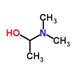
DMAE is a precursor to acetylcholine, which is one of the main memory-forming neurotransmitters in your brain. Higher acetylcholine levels have been linked with better performance on some memory tests.† As a nootropic, it is closely related to choline but due to a slightly different chemical structure it crosses the blood-brain barrier differently.†
Results: In rat experiments, DMAE p-Glu increased the extracellular levels of choline and acetylcholine in the medial prefrontal cortex, as assessed by intracerebral microdialysis, improved performance in a test of spatial memory, and reduced scopolamine-induced memory deficit in passive avoidance behavior. Clinical study results show that scopolamine induced a memory deficit and that DMAE p-Glu produced a significant positive effect on scores in the Buschke test, as well as a slight but significant difference on choice reaction time.
Psychopharmacology (Berl). 2009 Dec;207(2):201-12. doi: 10.1007/s00213-009-1648-7. Epub 2009 Sep 16.
Blin O1, Audebert C, Pitel S, Kaladjian A, Casse-Perrot C, Zaim M, Micallef J, Tisne-Versailles J, Sokoloff P, Chopin P, Marien M.
https://doi.org/10.1007/s00213-009-1648-7
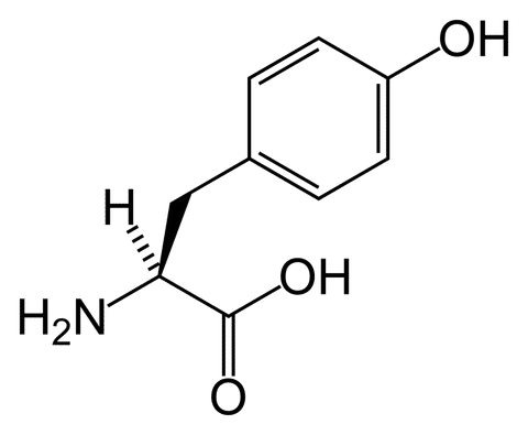
An amino acid considered a “building block” of protein. in some cases L-Tyrosine is also used to treat depression, anxiety, ADHD, cognitive disorders and treat chronic fatigue syndrome.†
Results: These results indicate that a free amino group is not required for recognition, provided that the modified amino group is able to take a positive charge. Steric factors appeared to be relatively unimportant.
J Drug Target. 2000;8(6):395-401.
Ohnishi T1, Maruyama T, Higashi S, Awazu S.
https://doi.org/10.3109/10611860008997915
Results: Therefore, these findings have two interesting implications for studies on TYR. First, the higher TH transcription activity associated with the T allele in the C-824T polymorphism could mean that low baseline DA individuals who carry this allele can benefit especially from TYR supplementation, as more TYR can be converted into DA. Second, the idea that the T allele protects against cognitive decline in schizophrenia could imply that it does so in old age as well. In that case, the effect of this polymorphism on TYR supplementation might be strongest in older individuals, as might be the case with the DRD2 gene. This possibility again underscores the need to keep age and age-related changes in neurophysiology in mind when investigating TYR.
Front Psychol. 2014 Oct 6;5:1101. doi: 10.3389/fpsyg.2014.01101. eCollection 2014.
Jongkees BJ1, Hommel B1, Colzato LS1.
https://doi.org/10.3389/fpsyg.2014.01101
Results: The findings indicate that loss of this intrinsic protective response pathway with age-dependent increase in oxidative stress may be a contributing factor for the susceptibility of the brain to age-associated neurologic disorders. DOI:
Neurobiol Aging. 2016 May;41:25-38. doi: 10.1016/j.neurobiolaging.2016.02.004. Epub 2016 Feb 12.
Rajagopal S1, Deb I1, Poddar R1, Paul S2.
10.1016/j.neurobiolaging.2016.02.004
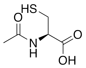
N-acetyl cysteine (sometimes refered to as NAC) comes from the amino acid L-cysteine, that has been known as a strong antioxidant and one of the few supplements to help combat hangover symptoms from Alcohol.†
Results: Molar ratios of the reduced and oxidized forms of GSH (GSH divided by glutathione disulfide, redox ratio) were also determined. The elimination phase half-life of NAC was approximately 34 min. Both labeled and unlabeled GSH in RBC were found to increase; however, the area under the curve above baseline (AUCb0-280 ) of labeled GSH was only 1% of the unlabeled form. These data indicate that NAC is not a direct precursor of GSH. In addition, NAC has prolonged effects in brain even when the drug has been eliminated from systemic circulation.
J Pharm Sci. 2015 Aug;104(8):2619-26. doi: 10.1002/jps.24482. Epub 2015 Jun 5.
Zhou J1,2, Coles LD1,2, Kartha RV1,2, Nash N3, Mishra U1,2, Lund TC3, Cloyd JC1,2.
https://doi.org/10.1002/jps.24482
Results: The current study evaluated a novel, cell permeant amide form of N-acetylcysteine (NACA), which has high permeability through cellular and mitochondrial membranes resulting in increased CNS bioavailability. Cortical tissue sparing, cognitive function and oxidative stress markers were assessed in rats treated with NACA, NAC, or vehicle following a TBI. At 15 days post-injury, animals treated with NACA demonstrated significant improvements in cognitive function and cortical tissue sparing compared to NAC or vehicle treated animals. NACA treatment also was shown to reduce oxidative damage (HNE levels) at 7 days post-injury. Mechanistically, post-injury NACA administration was demonstrated to maintain levels of mitochondrial glutathione and mitochondrial bioenergetics comparable to sham animals. Collectively these data provide a basic platform to consider NACA as a novel therapeutic agent for treatment of TBI.
Exp Neurol. 2014 Jul;257:106-13. doi: 10.1016/j.expneurol.2014.04.020. Epub 2014 May 1.
Pandya JD1, Readnower RD1, Patel SP2, Yonutas HM1, Pauly JR3, Goldstein GA4, Rabchevsky AG2, Sullivan PG5.
https://doi.org/10.1016/j.expneurol.2014.04.020
Results: Clozapine+NAC was more effective than clozapine alone in reversing SIR-induced PPI, mitochondrial, immune and DA changes. In conclusion, SIR induces mitochondrial and immune-inflammatory changes that underlie cortico-striatal DA perturbations and subsequent behavioural deficits, and responds to treatment with clozapine or NAC, with an additive effect following combination treatment. The data provides insight into the mechanisms that might underlie the utility of NAC as an adjunctive treatment in schizophrenia.
Brain Behav Immun. 2013 May;30:156-67. doi: 10.1016/j.bbi.2012.12.011. Epub 2012 Dec 25.
Möller M1, Du Preez JL, Viljoen FP, Berk M, Emsley R, Harvey BH.
https://doi.org/10.1016/j.bbi.2012.12.011
Results: These results suggest that redox-sensitive neutral SMase plays important role in SM turnover dysregulation in both the hippocampus and neocortex at old age and that the CR diet can prevent the age-dependent accumulation of ceramide mainly via neutral SMase targeting.
Arch Gerontol Geriatr. 2014 May-Jun;58(3):420-6. doi: 10.1016/j.archger.2013.12.005. Epub 2013 Dec 27.
Babenko NA1, Shakhova EG2.
https://doi.org/10.1016/j.archger.2013.12.005
Results: The combined taurine/NAC treatment also attenuated rotenone-induced reductions in aconitase activity suggesting the cytoprotection afforded by the combined treatment may be associated with anti-oxidative mechanisms. Together, our data suggest that a multi-targeted approach may yield new avenues of research exploring the utility of combining therapeutic agents with different mechanisms of actions at concentrations lower than previously tested and shown to be cytoprotective.
Brain Res. 2015 Oct 5;1622:409-13. doi: 10.1016/j.brainres.2015.06.041. Epub 2015 Jul 10.
Alkholifi FK1, Albers DS2.
https://doi.org/10.1016/j.brainres.2015.06.041

Created from the Brahmi plant which is native to the wetlands of India and has been a consistent part of traditional medicine for centuries. A 2013 study demonstrated that Bacopa Monniera extract has a potential to improve cognitive performance, particularly speed of attention by reducing choice reaction time.†
Results: Nine studies met the inclusion criteria using 518 subjects. Overall quality of all included trials was low risk of bias and quality of reported information was high. Meta-analysis of 437 eligible subjects showed improved cognition by shortened Trail B test (-17.9 ms; 95% CI -24.6 to -11.2; p<0.001) and decreased choice reaction time (10.6 ms; 95% CI -12.1 to -9.2; p<0.001).
J Ethnopharmacol. 2014;151(1):528-35. doi: 10.1016/j.jep.2013.11.008. Epub 2013 Nov 16.
Kongkeaw C1, Dilokthornsakul P2, Thanarangsarit P3, Limpeanchob N3, Norman Scholfield C4.
https://doi.org/10.1016/j.jep.2013.11.008
Results: Outcome measures included cognitive outcomes from the MTF, with mood and salivary cortisol measured before and after each completion of the MTF. Change from baseline scores indicated positive cognitive effects, notably at both 1 h post and 2 h post BM consumption on the Letter Search and Stroop tasks, suggesting an earlier nootropic effect of BM than previously investigated. There were also some positive mood effects and reduction in cortisol levels, pointing to a physiological mechanism for stress reduction associated with BM consumption. It was concluded that acute BM supplementation produced some adaptogenic and nootropic effects that need to be replicated in a larger sample and in isolation from stressful cognitive tests in order to quantify the magnitude of these effects.
Phytother Res. 2014 Apr;28(4):551-9. doi: 10.1002/ptr.5029. Epub 2013 Jun 21.
Benson S1, Downey LA, Stough C, Wetherell M, Zangara A, Scholey A.
https://doi.org/10.1002/ptr.50
Results: Bacopa monnieri extract improved the escape latency time (p<.01 in morris water maze test. moreover the reduction of neurons and cholinergic neuron densities were also mitigated.>
J Ethnopharmacol. 2010 Jan 8;127(1):26-31. doi: 10.1016/j.jep.2009.09.056. Epub 2009 Oct 4.
Uabundit N1, Wattanathorn J, Mucimapura S, Ingkaninan K.
https://doi.org/10.1016/j.jep.2009.09.056
Results: These findings imply a newer potential role of Bacopa monnieri in the clinical management of opioid withdrawal induced depression which can be attributed to Bacoside A3.
Phytother Res. 2014 Jun;28(6):937-9. doi: 10.1002/ptr.5081. Epub 2013 Nov 14.
Rauf K1, Subhan F, Abbas M, Ali SM, Ali G, Ashfaq M, Abbas G.
https://doi.org/10.1002/ptr.5081
Results: The findings showed upregulated expression of NR1 and decreased binding of REST/NRSF to NR1 promoter after postnatal exposure of PBDE-209. Interestingly, supplementation with CDRI-08 significantly restored the expression of NR1 and binding of REST/NRSF to NR1 promoter near to the control value at the dose of 120 mg/kg. In conclusion, the results suggest that CDRI-08 possibly acts on glutamatergic system through expression and regulation of NR1 and may restore memory, impaired by PBDE-209 as reported in our previous study.
Evid Based Complement Alternat Med. 2015;2015:403840. doi: 10.1155/2015/403840. Epub 2015 Aug 27.
Verma P1, Gupta RK2, Gandhi BS1, Singh P1.
http://dx.doi.org/10.1155/2015/403840
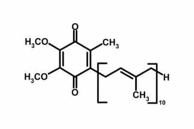
COQ10: Co-Enzyme Q10 is a compound present in mitochondria of every cell in the body. the results show that lower COQ10 plays a role in the pathophysiology of depression and in particular in TRD and CFS accompanying depression.† COQ10 supplementation also has been known to promote slower brain deterioration, as seen in a study published in the 2002 “archives of neurology.” †
Results: These results indicate that mitochondrial oxygen consumption in the brain decreases in aged male mice. Furthermore, these results suggest that exogenous CoQ10 restores mitochondrial oxygen use to levels equivalent to those observed in young mice.
Mech Ageing Dev. 2013 Nov-Dec;134(11-12):580-6. doi: 10.1016/j.mad.2013.11.010. Epub 2013 Dec 11.
Takahashi K1, Takahashi M2.
https://doi.org/10.1016/j.mad.2013.11.010
Results: The findings also support the hypothesis that brain energy impairment is involved in the pathophysiology of depression and enhancing mitochondrial function could provide an opportunity for development of a potentially more efficient drug therapy for depression.
Behav Pharmacol. 2013 Oct;24(7):552-60. doi: 10.1097/FBP.0b013e3283654029.
Aboul-Fotouh S1.
https://doi:10.1097/FBP.0b013e3283654029
Results: Our results demonstrate that CoQ10 is a robust neuroprotective agent against ischemia-reperfusion brain injury in rats, improving both functional and morphological indices of brain damage.
J Cardiovasc Pharmacol. 2016 Feb;67(2):103-9. doi: 10.1097/FJC.0000000000000320.
Belousova M1, Tokareva OG, Gorodetskaya E, Kalenikova EI, Medvedev OS.
https://doi:10.1097/FJC.0000000000000320
Results: These results demonstrate the beneficial effects of CoQ(10) against organophosphate-induced cognitive impairments and hippocampal neuronal degeneration.
Neurotox Res. 2012 May;21(4):345-57. doi: 10.1007/s12640-011-9289-0. Epub 2011 Nov 15.
Binukumar BK1, Gupta N, Sunkaria A, Kandimalla R, Wani WY, Sharma DR, Bal A, Gill KD.
https://link.springer.com/article/10.1007%2Fs12640-011-9289-0
Results: In the biochemical tests, tissue malondialdehyde (MDA) levels were significantly higher in the traumatic brain-injury group compared to the sham group (p < 0.05). Administration of CoQ₁₀ after trauma was shown to be protective because it significantly lowered the increased MDA levels (p < 0.05). Comparing the superoxide dismutase (SOD) levels of the four groups, trauma + CoQ₁₀ group had SOD levels ranging between those of sham group and traumatic brain-injury group, and no statistically significant increase was detected. Histopathological results showed a statistically significant difference between the CoQ₁₀ and the other trauma-subjected groups with reference to vascular congestion, neuronal loss, nuclear pyknosis, nuclear hyperchromasia, cytoplasmic eosinophilia, and axonal edema (p < 0.05).
BMC Neurosci. 2011 Jul 29;12:75. doi: 10.1186/1471-2202-12-75.
Kalayci M1, Unal MM, Gul S, Acikgoz S, Kandemir N, Hanci V, Edebali N, Acikgoz B.
https://doi.org/10.1186/1471-2202-12-75
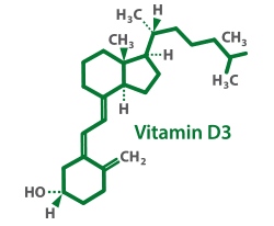
Vitamin D acts an immune system modulator and can be produced by the body using sunlight. present findings signify the crucial role of Vitamin D on the basic synaptic transmission and synaptic plasticity.† Due to weather changes, deficiency is common during the winter months. A recent study suggests that Vitamin D(3) supplementation during the winter may reduce the incidence of flu by 40%†
Results: These findings indicate that, vitamin D has anti-epileptic, cognitive improving and antioxidant effects, on its own and enhance the effects of lamotrigine, in a chronic model of epileptic seizures. Thus, vitamin D supplementation may be a useful addition to antiepileptic drugs improving seizure control and cognitive function in patients with epilepsy.
Brain Res. 2017 Oct 15;1673:78-85. doi: 10.1016/j.brainres.2017.08.011. Epub 2017 Aug 15.
Abdel-Wahab AF1, Afify MA2, Mahfouz AM2, Shahzad N2, Bamagous GA2, Al Ghamdi SS2.
https://doi.org/10.1016/j.brainres.2017.08.011
Results: Baseline ACC GABA/creatine (Cr) was lower in BSD than in TD (F[1,31]=8.91, p=0.007). Following an 8 week Vitamin D3 supplementation, in BSD patients, there was a significant decrease in YMRS scores (t=-3.66, p=0.002, df=15) and Children’s Depression Rating Scale (CDRS) scores (t=-2.93, p=0.01, df=15); and a significant increase in ACC GABA (t=3.18, p=0.007, df=14).
J Child Adolesc Psychopharmacol. 2015 Jun;25(5):415-24. doi: 10.1089/cap.2014.0110.
Sikoglu EM1,2,3, Navarro AA1,2,3,4, Starr D2,3, Dvir Y2,3, Nwosu BU5, Czerniak SM1, Rogan RC1, Castro MC2,3, Edden RA6,7, Frazier JA2,3,5, Moore CM1,2,3,8.
https://doi.org/10.1016/j.brainres.2017.08.011
Results: The present findings demonstrate the potential effect of vitamin D3 supplementation on cognitive function in diabetic animals. It is possible that this effect is mediated through enhancing the prefrontal cortex cholinergic transmission.
Behav Brain Res. 2015;287:156-62. doi: 10.1016/j.bbr.2015.03.050. Epub 2015 Mar 30.
Alrefaie Z1, Alhayani A2.
https://doi.org/10.1016/j.bbr.2015.03.050
Results: In summary, there is growing evidence that low prenatal levels of 1,25(OH)(2)D(3) can influence critical components of orderly brain development. It remains to be seen if these processes are of clinical relevance in humans, but in light of the high rates of hypovitaminosis D in pregnant women and neonates, this area warrants further scrutiny.
J Steroid Biochem Mol Biol. 2004 May;89-90(1-5):557-60.
McGrath JJ1, Féron FP, Burne TH, Mackay-Sim A, Eyles DW.
https://doi.org/10.1016/j.bbr.2015.03.050
Results: Our results confirm that vitamin D(3) regulates these processes in the developing brain at both cellular and molecular levels. Compared to control animals, the embryos and pups from vitamin D(3) depleted mothers had significantly less apoptotic cells, this finding being most pronounced at birth. Additionally, there were significantly more mitotic cells but this was not associated with any particular developmental period. Targeted gene arrays specific for apoptosis and cell cycle genes confirmed a pattern of transcription deregulation in the deplete group consistent with the known properties of vitamin D(3). While most current vitamin D(3) research is focussed on the pro-apoptotic and pro differentiating properties of vitamin D(3) as adjuncts for the treatment of cancers, our findings highlight the important role that this hormone plays in normal development via these same properties specifically in the brain.
Brain Res Dev Brain Res. 2004 Oct 15;153(1):61-8.
Ko P1, Burkert R, McGrath J, Eyles D.
https://doi.org/10.1016/j.devbrainres.2004.07.013

MethylCobalamin is more Bio-Available and expensive form of B12. The vitamin plays a key role in the normal functioning of the brain and nervous system.†
Results: The results of the present study showed that at the level of the dorsal hippocampus, vitamin B12 modulated orofacial pain through a mu-opioid receptor mechanism. In addition, vitamin B12 contributed to hippocampal cholinergic system in processing of memory. Moreover, cholinergic and opioid systems may be involved in improving effect of vitamin B12 on pain-induced memory impairment.
Physiol Behav. 2017 Mar 1;170:68-77. doi: 10.1016/j.physbeh.2016.12.020. Epub 2016 Dec 18
Erfanparast A1, Tamaddonfard E2, Nemati S2.
https://doi.org/10.1016/j.physbeh.2016.12.020
Results: Thus our findings reveal a previously unrecognized decrease in brain vitamin B12 status across the lifespan that may reflect an adaptation to increasing antioxidant demand, while accelerated deficits due to GSH deficiency may contribute to neurodevelopmental and neuropsychiatric disorders.
PLoS One. 2016 Jan 22;11(1):e0146797. doi: 10.1371/journal.pone.0146797. eCollection 2016.
Zhang Y1, Hodgson NW1,2, Trivedi MS1,3, Abdolmaleky HM4, Fournier M5, Cuenod M5, Do KQ5, Deth RC1,3.
https://doi.org/10.1016/j.physbeh.2016.12.020
Results: Thereby, it is important to treat vitamin B12 deficiency with a combination of MeCbl and AdCbl or hydroxocobalamin or Cbl. Regarding the route, it has been proved that the oral route is comparable to the intramuscular route for rectifying vitamin B12 deficiency.
Eur J Clin Nutr. 2015 Jan;69(1):1-2. doi: 10.1038/ejcn.2014.165. Epub 2014 Aug 13.
Thakkar K1, Billa G2.
https://doi.org/10.1038/ejcn.2014.165
Result: Concentrations of all vitamin B12-related markers, but not serum vitamin B12 itself, were associated with global cognitive function and with total brain volume. Methylmalonate levels were associated with poorer episodic memory and perceptual speed, and cystathionine and 2-methylcitrate with poorer episodic and semantic memory. Homocysteine concentrations were associated with decreased total brain volume. The homocysteine-global cognition effect was modified and no longer statistically significant with adjustment for white matter volume or cerebral infarcts. The methylmalonate-global cognition effect was modified and no longer significant with adjustment for total brain volume.
Neurology. 2011 Sep 27;77(13):1276-82. doi: 10.1212/WNL.0b013e3182315a33.
Tangney CC1, Aggarwal NT, Li H, Wilson RS, Decarli C, Evans DA, Morris MC.
https://doi.org/10.1038/ejcn.2014.165
Results: ReHo values in patients with vitamin B12 deficiency were significantly lower than controls in the entire cerebrum and the brain networks associated with cognition control, i.e., default mode, cingulo-opercular and fronto-parietal network. There was no significant difference using LFO and it did not show significant correlations with NPT scores. ReHo showed significant correlation with NPT scores. All the 6 patients showed increase in ReHo after replacement therapy.
Magn Reson Imaging. 2016 Feb;34(2):191-6. doi: 10.1016/j.mri.2015.10.026. Epub 2015 Oct 31.
Gupta L1, Gupta RK2, Gupta PK3, Malhotra HS4, Saha I5, Garg RK4.
https://doi.org/10.1016/j.mri.2015.10.026
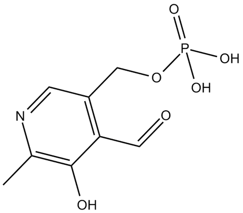
Pyridoxal-5-Phosphate, or P5P as it is commonly known, is a more expensive and active form of vitamin B6. P5P is a required coenzyme for the biosynthesis of several neurotransmitters, including GABA, dopamine, norepinephrine, and serotonin. Some studies show P5P increasing mood, memory, performance and mental effort.†
Results: From a medicinal chemistry perspective our results demonstrate that the issue of inhibitor specificity for highly conserved PLP-dependent enzymes could be successfully addressed.
J Med Chem. 2010 Aug 12;53(15):5684-9. doi: 10.1021/jm100464k.
Rossi F1, Valentina C, Garavaglia S, Sathyasaikumar KV, Schwarcz R, Kojima S, Okuwaki K, Ono S, Kajii Y, Rizzi M.
https://DOI:10.1021/jm100464k
Results: We hypothesize that both pyridoxal kinase and antiquitin 1 oxidation could result in decreased pyridoxal 5-phosphate availability necessary as a cofactor in transaminations, synthesis of glutathione, and synthesis of GABA and dopamine, two neurotransmitters that play a key role in HD pathology.
Free Radic Biol Med. 2010 Aug 15;49(4):612-21. doi: 10.1016/j.freeradbiomed.2010.05.016.
Sorolla MA1, Rodríguez-Colman MJ, Tamarit J, Ortega Z, Lucas JJ, Ferrer I, Ros J, Cabiscol E.
https://doi.org/10.1016/j.freeradbiomed.2010.05.016
Results: These results indicate that vitamin B-6 deficiency affects one or more structures of the brainstem that generate the later parts of the BAEP. The finding of prolonged interwave intervals in vitamin B-6-deficient animals is consistent with slowed axonal conduction velocity secondary to defective myelination. Recording BAEP provided a noninvasive means of detecting effects of vitamin B-6 deficiency on specific parts of the central nervous system.
J Nutr. 1993 Jan;123(1):20-6.
Buckmaster PS1, Holliday TA, Bai SC, Rogers QR.
https://doi.org/10.1093/jn/123.1.20
Results: Norepinephrine in the raphe nucleus followed a similar biphasic pattern. Excess dietary pyridoxine affected brain and serum concentrations of some amino acids and binding properties of cortical serotonin receptors in a biphasic pattern over the range of concentrations fed in this study.
J Nutr. 1998 Oct;128(10):1829-35.
Schaeffer MC1, Gretz D, Gietzen DW, Rogers QR.
https://doi.org/10.1093/jn/128.10.1829
Results: Brain weight and volume of neocortex were not changed significantly by the maternal restrictions imposed. However, each restriction adversely affected neurogenesis and neuron longevity of the neocortex and when expressed as percent reduction from control, neuron longevity was affected more severely than neurogenesis.
J Nutr. 1987 Jun;117(6):1045-52.
Groziak SM, Kirksey A.
https://doi.org/10.1093/jn/117.6.1045

Also known as Niacin. this vitamin is known as a key Co-Enzyme in the production of cellular energy. Data from research suggests Vitamin B3 may be beneficial in preserving and enhancing neuroconitive function.†
Results: Recent work investigating the effects of nicotinamide riboside in yeast and mammals established that it is metabolized by at least two types of metabolic pathways. The first of these is degradative and produces nicotinamide. The second pathway involves kinases called nicotinamide riboside kinases (Nrk1 and Nrk2, in humans). The likely involvement of the kinase pathway is implicated in the unique effects of nicotinamide riboside in raising tissue NAD concentrations in rodents and for potent effects in eliciting insulin sensitivity, mitochondrial biogenesis, and enhancement of sirtuin functions. Additional studies with nicotinamide riboside in models of Alzheimer’s disease indicate bioavailability to brain and protective effects, likely by stimulation of brain NAD synthesis.
Curr Opin Clin Nutr Metab Care. 2013 Nov;16(6):657-61. doi: 10.1097/MCO.0b013e32836510c0.
Chi Y1, Sauve AA.
https://DOI:10.1097/MCO.0b013e32836510c0
Results: The protective effect observed, at biologically relevant concentrations, with nicotinamide was more than that of the endogenous antioxidants ascorbic acid and alpha-tocopherol. Hence our studies suggest that nicotinamide (vitamin B3) can be considered as a potent antioxidant capable of protecting the cellular membranes in brain, which is highly susceptible to prooxidants, against oxidative damage induced by ROS.
Redox Rep. 1999;4(4):179-84.
Kamat JP1, Devasagayam TP.
https://doi.org/10.1089/089771503770802871
Results: These results indicate that B(3) administration significantly improved behavioral outcome following injury, reduced the size of the lesion, and reduced the expression of GFAP. The current findings suggest that B(3) may have therapeutic potential for the treatment of TBI.
J Neurotrauma. 2003 Nov;20(11):1189-99.
Hoane MR1, Akstulewicz SL, Toppen J.
https://doi.org/10.1089/089771503770802871
Results: Taken together, our findings demonstrate that NR is a better neuroprotective agent than NAD+ in excitotoxicity-induced AxD and that axonal protection involves defending intracellular NAD+ homeostasis.-Vaur, P., Brugg, B., Mericskay, M., Li, Z., Schmidt, M. S., Vivien, D., Orset, C., Jacotot, E., Brenner, C., Duplus, E. Nicotinamide riboside, a form of vitamin B3, protects against excitotoxicity-induced axonal degeneration.
FASEB J. 2017 Dec;31(12):5440-5452. doi: 10.1096/fj.201700221RR. Epub 2017 Aug 21.
Vaur P1, Brugg B1, Mericskay M1,2, Li Z1,3, Schmidt MS4, Vivien D5, Orset C5, Jacotot E1, Brenner C4, Duplus E6.
https://doi.org/10.1096/fj.201700221RR
Results: Data from animal and human interventional studies and epidemiological research suggests that nicotinamide may be beneficial in preserving and enhancing neurocognitive function. Discussion Nicotinamide is non-toxic, inexpensive and widely available, and interventional studies in humans, using supplemental doses of nicotinamide, are now warranted.
Nutr Neurosci. 2015 Jul;18(5):193-200. doi: 10.1179/1476830514Y.0000000112. Epub 2014 Feb 21.
Rennie G, Chen AC, Dhillon H, Vardy J, Damian DL.
https://doi.org/10.1179/1476830514Y.0000000112
† These statements have not been evaluated by the FDA. This product is not intended to diagnose, treat, cure, or prevent any disease.